- +91 - 90 6646 2996 | 90 6636 2996
- +91 - 40 - 2330 2996 | 40 - 2335 0696
- drraghuveerreddy3@gmail.com
Articular cartilage injuries
The treatment options for articular cartilage injuries
Some patients with an articular cartilage injury improve with conservative treatment. The treatment includes exercises, use of anti-inflammatory medications (NSAIDs). The exercise may include a program you can do at home or formal physical therapy. Depending on the extent of the damage, some patients get better with these treatments and do not require surgery. If patients do not get better with conservative therapy, or have a large articular cartilage lesion, surgery may be necessary.
Surgical treatment of articular cartilage injuries
The surgery for articular cartilage injuries depends on the extent of the damage. There are several surgical options, and which procedure is best depends on several factors. These factors include the patient´s age and activity level, the size of the lesion, and the chronicity (age) of the lesion.
Surgical options include
- Smoothing of the lesion and removing loose edges only (debridement)
- Techniques to stimulate scar cartilage to grow into the lesion (microfracture)
- Techniques to replace the lesion with new cartilage (osteochondral autografts, osteochondral allografts, or autologous chondrocyte implantation).
Debridement of the articular cartilage
Debridement is performed by using small arthroscopic instruments, such as a mechanical shaver, to smooth the cartilage edges. This is performed so that there are no loose edges to irritate the joint, and to prevent the area of damage from expanding.
The microfracture technique
Microfracture is a technique to attempt to repair damaged articular cartilage. Small holes are made in the denuded bone in order to allow blood and marrow healing elements into the area of missing cartilage. This technique allows scar cartilage (fibrocartilage) to fill the area where the cartilage is missing. This technique is easy to perform and can create good results in a lot of patients. However, since the cartilage is scar cartilage, it may not be as durable as other techniques to restore cartilage defects. Microfracture can be performed during an arthroscopy, and no other incision or surgery is needed.
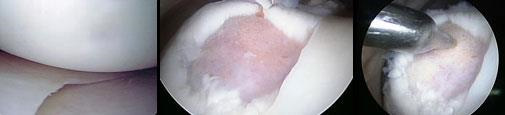
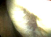
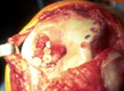
Normal healthy articular cartilage in the knee (left). A large cartilage defect in the knee joint surface (center). During microfracture, an awl is used to penetrate the defect (right)
Osteochondral Autograft (Mosaicplasty)
An osteochondral autograft is a technique to take a small piece of cartilage and bone from one area of the knee and put it in the area that the cartilage is missing. The cartilage is taken from an area in the knee that having minimal stress, so it is thought that patients do not have symptoms due to piece of cartilage loss.This technique can be very effective for small areas of cartilage damage in weight bearing areas. An osteochondral autograft can often be performed by arthroscopic techniques, but sometimes requires an open incision on the knee.
Autologous Chondrocyte Implantation (ACI)
Autologous chondrocyte implantation (ACI formerly referred to as autologous cartilage transplantation or ACT) is an approach that has been used to treat defined, symptomatic knee cartilage defects. ACI comprises a series of procedures.(This is a two staged procedure)
Cartilage Cell Therapy
- No Surgery
- No Medicine
- No Side Effects

Innovative cell therapy just for you!
Autologous Chondrocyte Implantation (ACI) is procedure that uses patients own cartilage tissue to treat defect area. Cartigrow cell therapy is a India's first ACI which is personalized, natural and curative treatment that regenerates the right type of cartilage. This enables the patient to perform all daily activities including sports. It helps in preventing total knee replacement in patients aged 18-65 years.
Indications
- Cartilage damage due to sports injury
- Early osteoarthritis
- Minimally invasive arthroscopic procedure
- Regeneration of hyaline-like cartilage
- Further progression of the disease is ceased
- Active normal life including sports
Benefits of CARTIGROW
Cartilage Cell Therapy Process
Biopsy
Step I:
In a minor Arthroscopic procedure, a punch biopsy of healthy cartilage along with subchondral bone will be picked up and transported in specially designed kit to the cell processing center. Temperature during transit is maintained.
Culture
Step II:
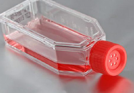
Cartilage making chondrocytes cells will be isolated and multiplied to obtain a dose of 48 million containing chondrocytes. A number of quality analysis tests are performed on the cell dose. The process takes about 4 weeks
Implantation
Step III:
In an open procedure that requires about 30 minutes, the defect area is prepared and the personalized dose of chondrocytes will be implanted in to the defect area. The cell-gel mixture takes the shape of the defect area and settles in a firm jelly within 8-10 minutes.
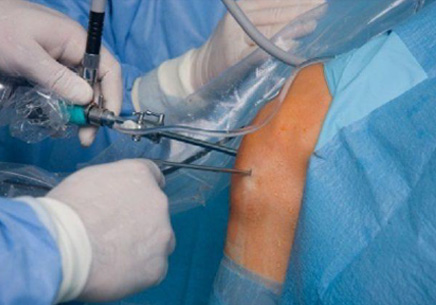
Bone Marrow Aspirate Concentrate Implantation
(This is a single stage procedure)
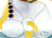
This is a minimally invasive procedure where in the fluid is taken from bone marrow. This fluid contains stem cells that can help the healing of some bone and joint conditions. These Stem cells help with bone healing, cartilage repair and new blood vessel growth, thus helping to treat various conditions like Bone marrow removal from the pelvis of a patient delayed union or nonunion of bone fractures, cartilage damage, osteonecrosis, chronic tendon problems or chronic wounds. This method is avoided in patients who have malignancy or infection.
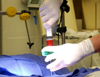
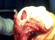
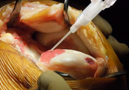
Bone marrow removal from the pelvis of a patient
Brief Description of Procedure
The procedure is done under local/ general anesthesia. A needle is used to remove bone marrow from within the bone.
Common site for the marrow aspirate is pelvis usually on the same side as that of foot or ankle procedure.
The sample of bone marrow is taken and then run down in a centrifuge , which seperates the cells, resulting in a high concentrated liquid, rich in stem cells.This high concentrated liquid is then injected directly into the cartilage surgery site.
Osteochondral Allograft Transplantation
An allograft is considered when the cartilage defect is much bigger that cannot be covered with an auto-graft. This procedure is typically done by an open incision. For this procedure, a tissue graft is taken from a cadaver donor, prepared and sterilized in a laboratory. Then the tissue is checked for any possible transimission of diseases. It is shaped to fit the exact contour of the defect and then press fit into place.