- +91 - 90 6646 2996 | 90 6636 2996
- +91 - 40 - 2330 2996 | 40 - 2335 0696
- drraghuveerreddy3@gmail.com
Calcific Tendinitis Treatment
Calcific tendinitis (or Tendonitis)
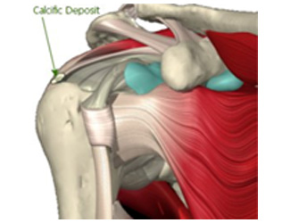
Calcific tendinitis is characterised by deposition of calcium in your rotator cuff. Calcium buildup in this area can restrict shoulder movements, as well as cause pain and discomfort.
Who is Effected
Calcific tendinitis is one of the most common cause of shoulder pain. It mostly effects people performing more overhead motions, such as heavy weight lifting, or sports like basketball or tennis. This condition is typically seen in adults between 40 and 60 years of age. Women are more commonly affected than men.
Symptoms
Though shoulder pain is the most common symptom in calcific tendinitis, about one-third of people may not experience any noticeable symptoms. Some may find that they're unable to move their arm, others may experience disturbed sleep, due to severe pain.
What Causes this Condition?
genetic predisposition
- Abnormal cell growth
- Abnormal thyroid activity
- Bodily production of anti-inflammatory agents
- Metabolic diseases such as diabetes
Diagnosis
After your physical examination, doctor may likely recommend imaging tests to look for any calcium deposits or other abnormalities.
- An X-ray can reveal larger deposits, and an ultrasound can help your doctor locate smaller deposits that the X-ray missed.
Treatment options
Most cases of calcific tendinitis can be treated conservatively. In mild cases, your doctor may recommend a mix of medication along with physiotherapy or a nonsurgical procedure.
Medication
Nonsteroidal anti-inflammatory drugs (NSAIDs) are considered to be the first line of treatment.
Nonsurgical procedures
In mild-to-moderate cases, we recommend one of the following procedures.
Extracorporeal shock-wave therapy (ESWT)
Radial shock-wave therapy (RSWT)
Therapeutic Ultrasound
Percutaneous needling: This therapy is more invasive than other nonsurgical methods. After injecting local anaesthesia to the area, a needle is used to make small holes in your skin. This will allow to manually remove the deposit. This may be done under ultrasound guidance to help the needle move into the correct position.
Surgery
About 10 percent of patients will require surgery to remove the calcium deposit.
An incision in the skin is made directly above the deposit's location and deposit is removed manually.
If arthroscopic surgery is performed, doctor will make a small incision and insert a tiny camera. The camera will guide the surgical tool in removal of the deposit.
Your recovery period will depend on the size, location, and number of calcium deposits.
Rehabilitation without surgery
The physiotherapist will teach you a series of gentle range-of-motion exercises to help improve movement in the affected shoulder. Exercises such as the pendulum, with slight swinging of the arm, are often prescribed at first. Over time, you'll work up to limited range-of-motion, isometric, and light weight-bearing exercises.
Rehabilitation after surgery
Recovery time after surgery varies from person to person. In some patients, full recovery may take three months or longer. Recovery from arthroscopic surgery is generally quicker compared to open surgery.
After either open or arthroscopic surgery, you are advised to wear a sling for a few days to support and protect the shoulder.
You should also attend physiotherapy sessions for 6 to 8 weeks. Physical therapy usually begins with some stretching and very limited range-of-motion exercises. You will typically progress to some light weight-bearing activity in about four weeks time.